Jul 01, 2023
Single Crystal Analysis by XtaLAB Synergy-ED
ED2022-01E
Innovative XtaLAB Synergy-ED for structural analysis of submicron crystals
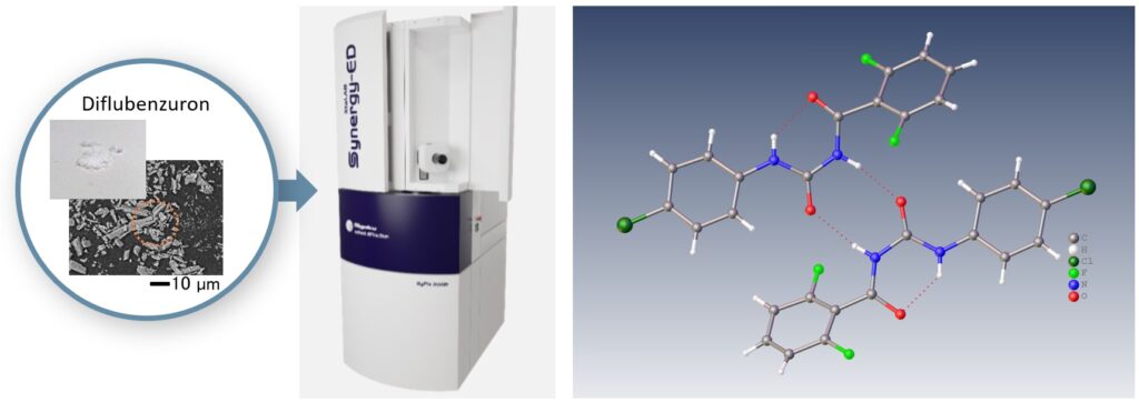
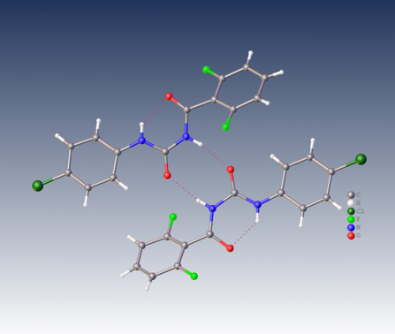
Right : Electron diffraction structure analysis of Diflubenzuron, XtaLAB Synergy-ED
XtaLAB Synergy-ED is a fully optimized electron diffractometer for microcrystals analysis, jointly developed by both companies: Rigaku Corporation and JEOL Ltd. The XtaLAB Synergy-ED features Rigaku’s high-speed and high-sensitivity photon-counting detector (HyPix-ED) and a state-of-the-art instrument control with a single crystal analysis software platform (CrysAlisPro for ED). The key characteristic of this product provides researchers an easy and efficient platform for electron crystallography. The right figure shows the result of structural analysis of a Diflubenzuron (C14H9ClF2N2O2) micro-crystal analyzed directly by XtaLAB Synergy-ED.
Sample preparation
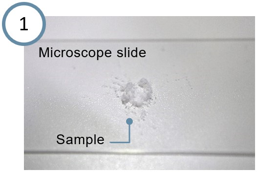
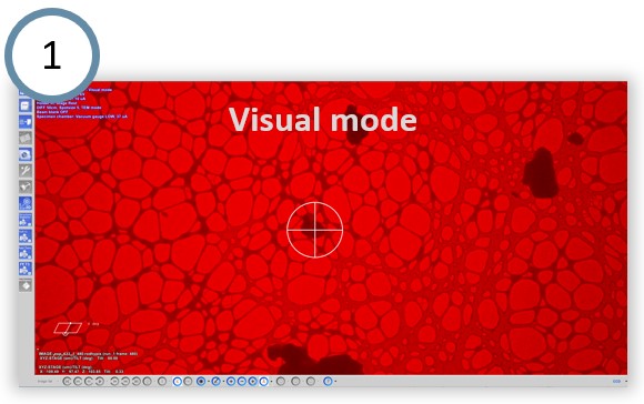
No sample preparation is required. XtaLAB synergy-ED uses a grid for TEM observation: load your sample directly onto a TEM grid as follows. If your particles are large, grind them with a microscope slide or spatula to a fine particle size.

3D ED/microED experiments
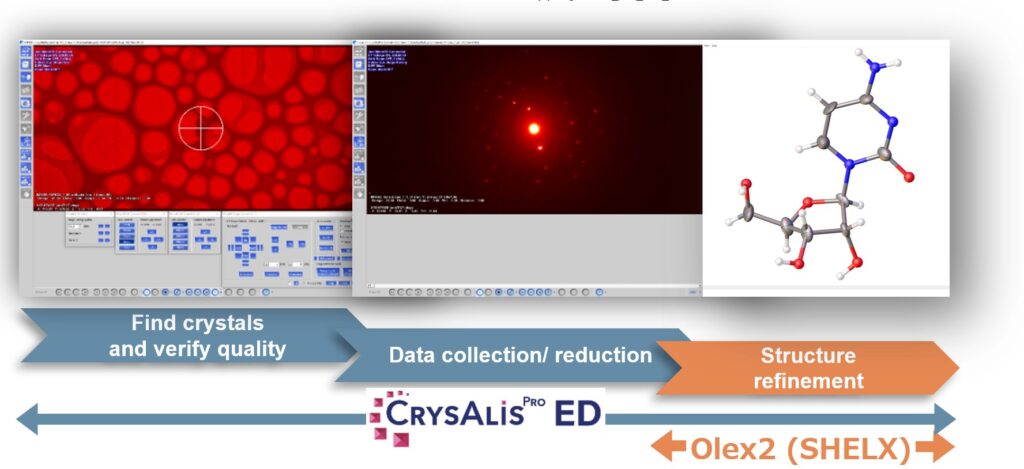
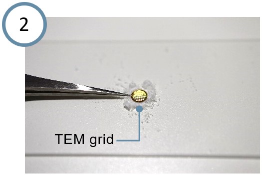
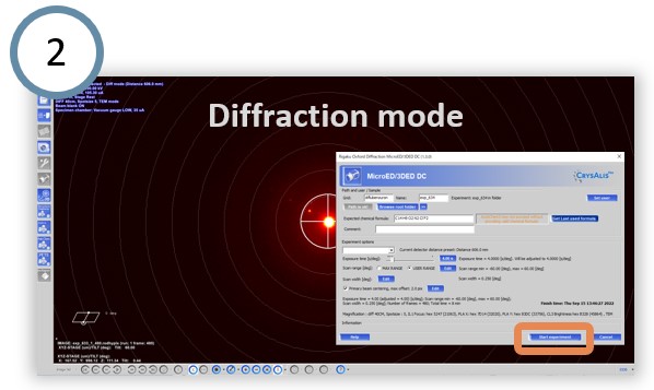
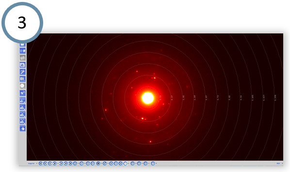
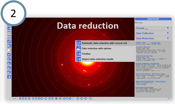
CrysAlisPro for ED allows easy switching between the visual mode and the diffraction mode. In the visual mode, click on an appropriate particle and the Diff.exp button in the MicroED Stage Control menu to switch into the diffraction mode. Then click the Start experiment button in the MicroED/3D ED DC window to start the electron diffraction observation automatically. The rotation range used for a typical measurement is −60 to +60 degrees.
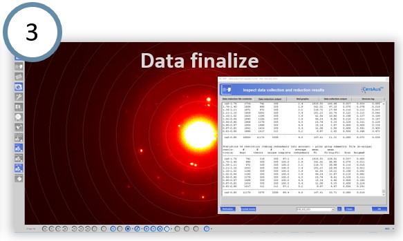
Structure analysis
In CrysAlisPro for ED, Autochem performs fully automatic structure determination and refinement during data collection. In addition, it is also possible to manually analyze the structure with efficient and effective functions and merge the dataset obtained from multiple crystals to improve the completeness of the data.

Structure refinement
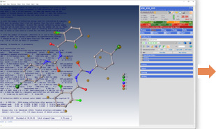
CrysAlisPro for ED allows seamlessly switching to Olex2. The intuitive user interface allows you to build and refine your three-dimensional molecular structure.
Solutions by field
- Others
Related products