Product
XtaLAB Synergy-ED, Platform for Electron Diffraction Structure Analysis without expertise
In recent years, there has been an increasing need for single crystal structural analysis of materials for which only very small crystals of less than a few hundred nanometers can be obtained. Such analysis was not possible with conventional technologies. Then, the discussion about an analytical method called 3D ED/MicroED became active and it began to be expected as a technique of structure analysis of very small crystals which is difficult to apply single crystal X-ray structure analysis. However, the conventional method using electron beams was not able to obtain enough data to perform single crystal structure analysis, as the analysis target substance was heavily damaged by electron beams. Furthermore, the sequence of steps required from data collection to analysis required for structure determination by electron diffraction required the assistance of experts of electron microscope and crystallographers, which compromised the speed and efficiency required at the research sites.
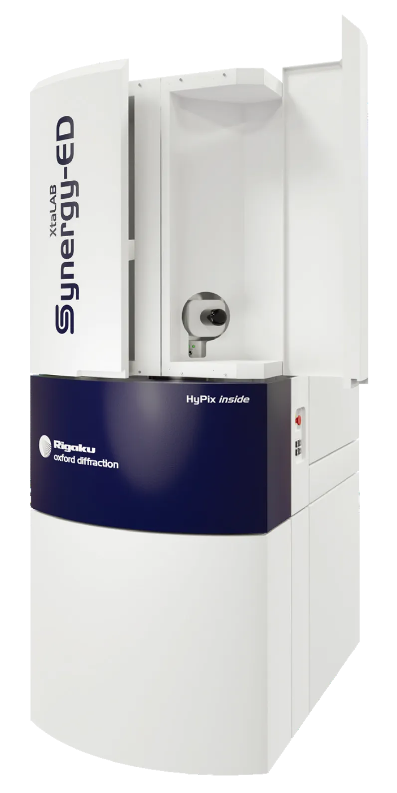
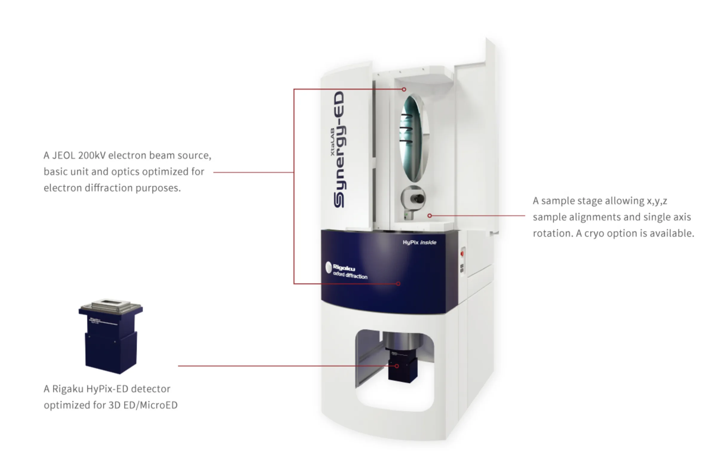
In order to resolve such an issue, RIGAKU and JEOL have jointly developed the electron diffractometer”XtaLAB Synergy-ED” which combines RIGAKU’s core technologies of single crystal structure analysis instruments with JEOL’s transmission electron microscopy technologies.
“XtaLAB Synergy-ED” performs a series of processes from data collection to analysis seamlessly, allowing for a prompt start of structure analysis using electron diffraction upon delivery. Thus, it is hoped for a contribution in not only with academia but also the latest research and development sites including new drug development and material development.
Strengths of XtaLAB Synergy-ED
In the past, to perform structure analysis using 3D ED/MicroED, it was necessary to obtain diffraction patterns by using the camera and software provided with the electron microscope for photographing and process the data using another crystallographic software.
Most cameras are dedicated to obtaining electron microscopic images and are not suitable to obtain high quality electron diffraction that are required to analyze a single crystal structure.
To overcome this situation, HyPix-ED which is a world-reputed direct exposure photon counting detector in single crystal X-ray structure analysis field ”HyPix” made applicable for electron measurement, and “CrysAlisPro for ED which is software for 3D ED/MicroED based on data measurement and processing software “CrysAlisPro”, were developed and installed in XtaLAB Synergy-ED.
Main Features of XtaLAB Synergy-ED
-

Seamless workflow from data measurement to crystal structure determination
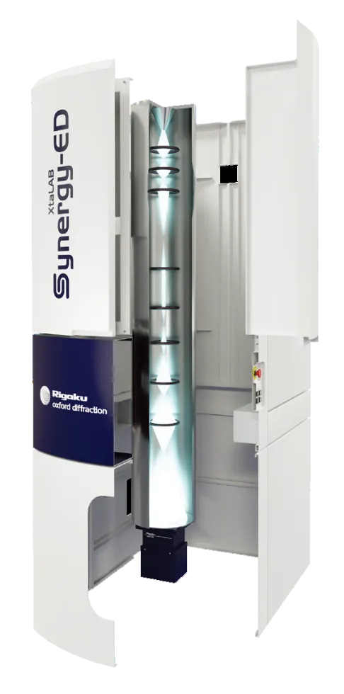
XtaLAB Synergy-ED has realized a seamless flow from measurement to analysis, which can visualize the 3D structure of atoms/molecules using electron beams. A series of operations from sample selection, diffraction data acquisition, to structure analysis can be easily performed by CrysAlisPro for ED. This is the outcome of combinations of RIGAKU’s ultra-high sensitivity high speed detector”HyPix-ED” and dedicated single crystal structure analysis software “CrysAlisPro for ED” with JEOL’s technologies accumulated through transmission electron microscopes.
-
Structure determination is possible for small crystals of several hundreds nanometers or less, by utilizing the advantage of electron diffraction that can measure nano-sized crystals.
Single crystal electron diffraction analysis is not a substitute for single crystal X-ray structure analysis. It is an analytical technique suitable for determination of a small crystal structure which is difficult to measure using X-ray diffraction. This is possible because the interaction between an electron beam and atoms is thousands to tens of thousands stronger than with X-rays. The strong interaction is both an advantage and a disadvantage at the same time. One of the disadvantages is that there is an upper limit in the size (thickness) of crystals that can be measured. The size (thickness) of crystal measurable with 3D ED/MicroED is less than the limit measurable with X-rays. In terms of measurable crystal size, there is no overlapping between the two methods. In fact, they are complementary analysis methods.
-
Speedy and simpler measurement due to development of electron diffractometer specialized for electron diffraction
3D ED/MicroED using a general-purpose transmission electron microscope requires knowledge and know-how of electron microscopy for setting up for transmission image acquisition and electron diffraction. The operation itself is often difficult for users who are not familiar with electron microscopy. For this reason, the XtaLAB Synergy-ED is specialized in functions as an electron diffractometer rather than an electron microscope, and integrates the CrysAlisPro for ED. This is optimized for electron diffraction, based on the CrysAlisPro, which has an established reputation for X-ray diffraction. The system allows the user to perform everything from crystal selection to data measurement, data processing and structural analysis in a single software package.Researchers who have used single crystal X-ray structure analysis can start analyzing the structure of a substance for which only nano-sized crystals are available immediately after installation.
-

Researchers who have usted single crystal X-ray structuer analysis, can immediately start using it. No need for the expertise of electron microscopy.
By integrating the flow from selection of measurement samples (nanocrystals) to data collection and analysis, electron diffraction can be easily used by non-specialists who lack the expertise in electron microscopy and crystallography that is conventionally required.
Structure analysis using XtaLAB Synergy-ED
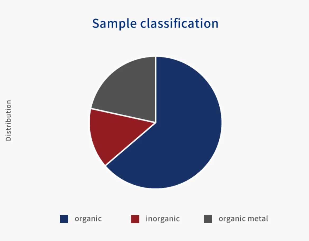
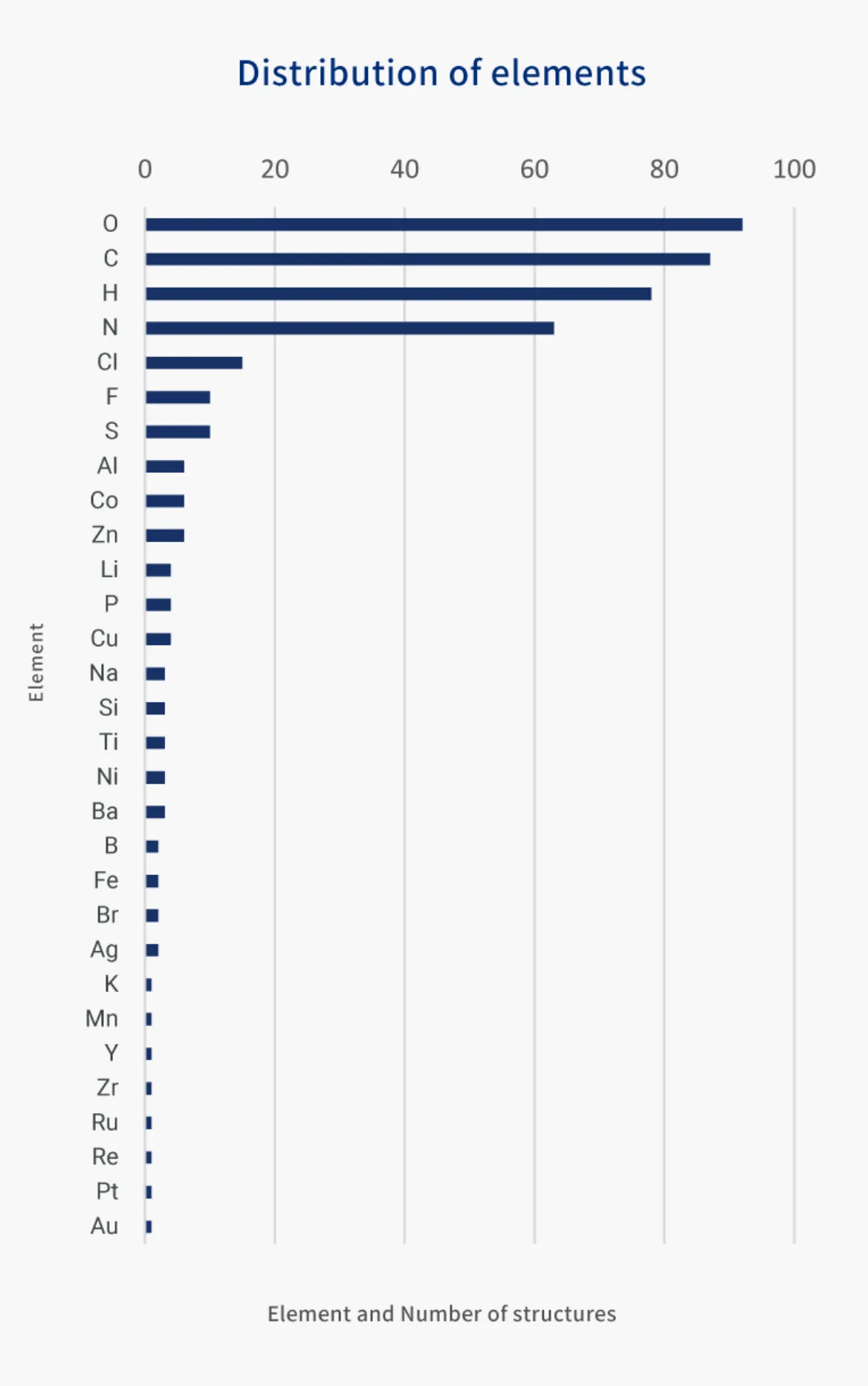
Since its launch, XtaLAB Synergy-ED has determined one structure after another. More than 100 structures have already been determined by electron diffraction from nanocrystals. A wide range of samples have been analyzed, from MOFs and organic molecules to structures containing as many as 1,600 atoms per unit lattice.
Note: Refinement only based on kinetic theory
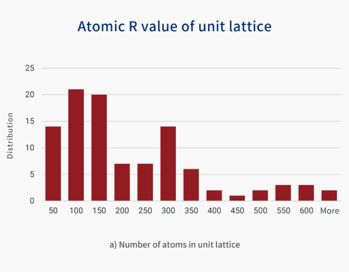
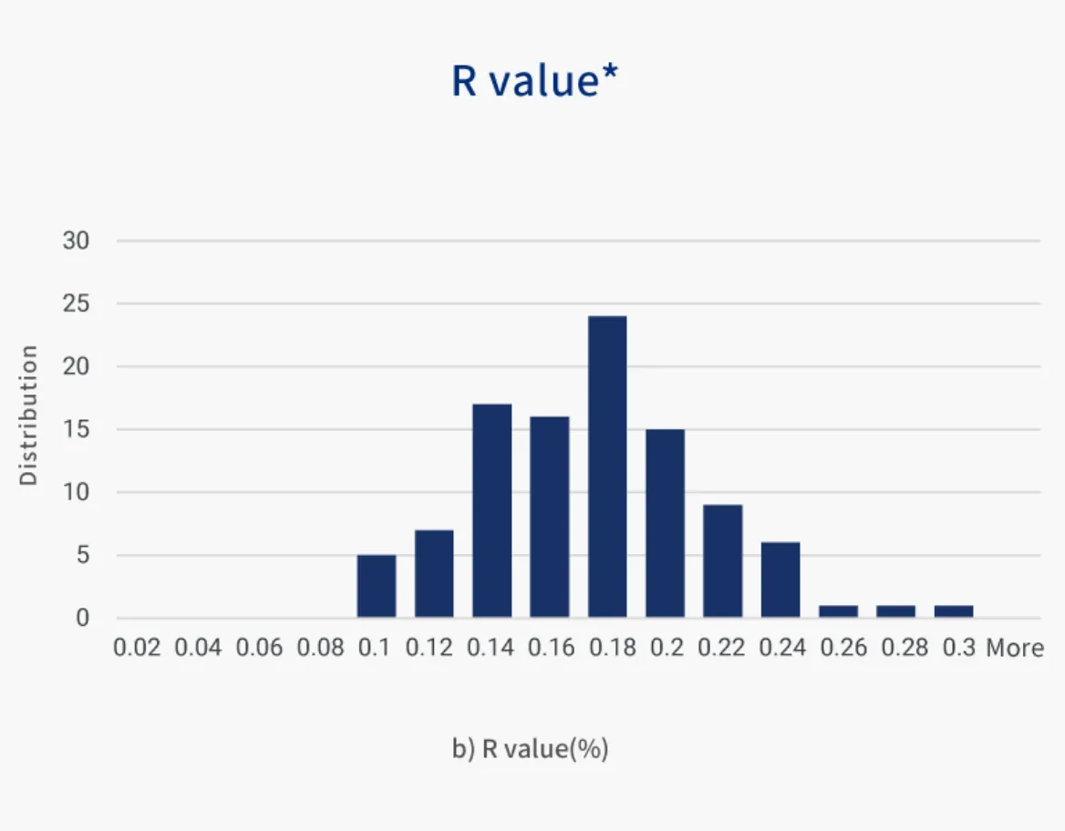
Figure 1: a) number of atoms per unit lattice, b)R value obtained by refining based on kinetic theory for the structures that have been analyzed.
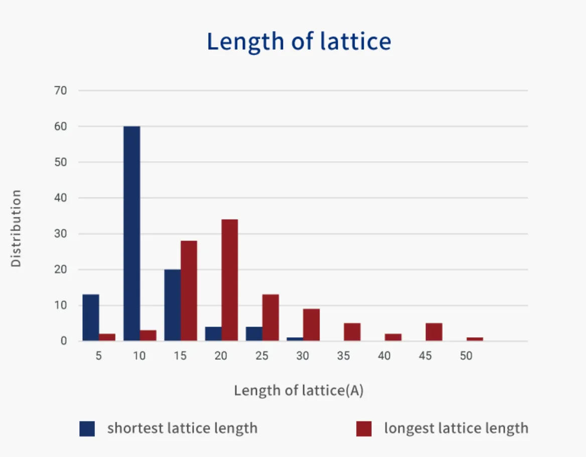
XtaLAB Synergy-ED supports very small crystals of a sample
that has short and long lattices, various compositions
Hardware
Specifications and features
| Product name | XtaLAB Synergy-ED |
|---|---|
| Technique | Single crystal electron diffraction |
| Core attributes | Dedicated electron diffractometer widh hybrid pixel array detector,rotation axis, and CrysAlisPro-ED,a complete integrated software platform. |
| Detector | High-speed,high-sensitivity hybrid pixel array detector,HyPix-ED |
| Source | 200 kV |
| Goniometer | Single rotation axis |
| Options | Various low temperature devices |
| Computer | External PC, MS Windows®︎ OS |