Solutions
Single Crystal
Structure Anlaysis
When x-rays or neutrons are illuminated onto a crystal that have atoms and molecules arranged periodically, they are scattered in the crystal, causing diffraction phenomena. The obtained diffraction patterns are the reflection of the arrangement of atoms and molecules in the crystal. Analyzing the obtained diffraction patterns in details allows us to obtain the structures of atoms and molecules of the measured material.
Crystals are largely divided into single crystals that are composed of single ordered structures and polycrystals that are the structures composed of multiple single crystals. Structural analysis by diffraction for single crystals is highly accurate and used in a wide range of samples from inorganic materials to biological materials such as proteins.
What is electron diffraction?
Electron diffraction is a method to obtain the arrangement of atoms (molecules) of a material, by using the diffraction phenomenon caused by irradiating electron beams to a material(crystal). In order to obtain diffraction patterns from a crystal, illumination of an x-ray or neutrons is required. The diffraction patterns also can be obtained by irradiating electrons that are charged particles.
In particular, electrons are negatively charged particles, so they can be easily bent in magnetic/electric fields. This property was utilized to develop electron microscopes. Electron microscopes are utilized in many researches since they can freely control magnification due to the combination of magnetic field lenses and the resolution is also high. Electron microscopes also have functions of observing electron diffraction in addition to the observation of an magnified image of the target. It is possible for electron microscopes to obtain diffraction patterns from the desired area of observation target material by combining these functions. In addition, the interaction between electrons and material is much larger than that of X-rays. Therefore, electron diffractions can be easily obtained even for crystals smaller than 1um.
3D ED/MicroED
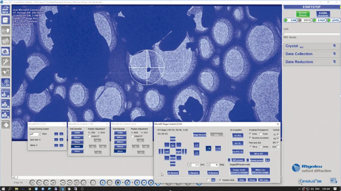
A revolutionary technique for obtaining 3D molecule structures from nanocrystalline materials. This method allows us to obtain diffraction patterns of small single crystals by illuminating electron beams. By continuously tilting the target single crystal against the illuminating electron beams, the tilted electron diffraction series can be obtained around a certain crystal axis. Use of the obtained diffraction patterns in the same manner as the analytical method “direct method” used with the single crystal structure analysis using X-ray, an analysis of the crystal structure is possible.
While an X-ray needs to use a relatively large single crystal of more than 1μ, 3D ED/MicroED using electron diffraction, is possible for small single crystals of less than 1μ, it is especially useful for a sample where a large single crystal is not available.
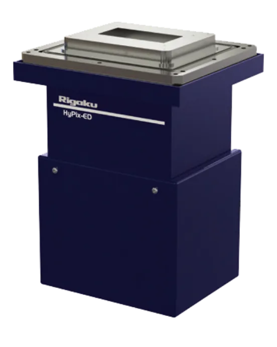
HyPix-ED
A hybrid type high performance pixel detector allowing direct detection of electrons. Capable of higher performance in sensitivity, noise level, measuring speed, than a conventional scintillator type detector that detects electrons.
HyPix-ED can measure the weak intensity of diffraction electrons and has a very high dynamic range required.
In electron diffraction, there are two types of electrons:
Transmission electrons, which transmit the sample without diffraction. Diffracted electrons, which are very weak compared to transmission electrons.
Low Molecule Crystal Structure Analysis
Analysis of a low molecule crystal is applicable for compounds that are chemical and biological interest, such as new synthetic chemical substance, catalysts, pharmaceuticals, and natural products. In particular, while it is frequently used to distinguish natural products and products of synthetic chemical experiments, it is also used for researches including molecular shape analysis, molecular interaction, and absolute configuration.
This technique, sometimes called chemical crystallography, clearly determines the 3D structure of molecules by using a single crystal X-ray diffraction (SXRD). This technique is to study mainly chemical property matters using crystallography, allowing for molecular dimension measurements with high accuracy and precision, which is not possible with other methods.
Drug Design
Since XtaLAB Synergy-ED enables analysis of very small crystals, structure of substances where crystals are hard to grow can be elucidated. Moreover, many crystals can be set to the instrument at once, and the evaluation of crystal quality is easy, allowing measurement to be performed with a selection of crystals. Therefore, it is not needed to replace the crystal for every measurement, reducing measurement steps. These XtaLAB Synergy-ED features bring about great benefits to drug discovery research.
The scenes of utilization are explained in terms of 1.confirmation of planar structure, 2.determination of absolute arrangement, 3.elucidation of crystal polymorphism.
1.Confirmation of planar structure
In drug discovery research, it is often necessary to elucidate or confirm the planar structure of a substance. For example, for drug metabolite, decomposition product, impurities, crystal structure is normally not analyzed because of the limited amount of available samples, and structures are estimated by mass analysis only. In case a more precise structure analysis is needed, the target substance is separated by HPLC and the structure is analyzed by NMR. But because of NMR sensitivity, enormous efforts are required to obtain as many samples as needed for NMR analysis. In some cases, the amount of sample required for NMR analysis cannot be obtained because the absolute amount is too small. In such a case, crystallization of small sample amount and XtaLAB Synergy-ED measurement is a new option. The feature of analysis capability with small sample volume is also useful for structural analyses of trace amount of natural products and synthetic compounds with small sample volumes. In addition, similar to X-ray crystal structure analysis, its use can be expected for structure analysis of compounds consisting of a few hydrogens which is difficult with the NMR analysis.
2.Determination of absolute arrangement
Determination of absolute arrangement is very important in drug discovery research, as it can impact pharmacological activity greatly. With XtaLAB Synergy-ED, determination of absolute arrangement considering multiple dispersion is reported, in addition to the estimation of absolute arrangement by salt formation with a chiral compound. For a substance in which absolute arrangement is not considered because of sample volume, XtaLAB Synergy-ED can provide absolute arrangement information, allowing us to obtain new knowledge.
3.Elucidation of crystal polymorphism
XtaLAB Synergy-ED can obtain coordinates information of each atom, revealing crystal polymorphism in addition to 3D structures of molecules. The difference of crystal polymorphism affects stability and solubility, and ultimately changes in vivo pharmacokinetics and medicinal benefits, and toxicity. Therefore, it is important to elucidate and understand the crystal polymorphism. In addition, the difference of crystal polymorphism of drug can cause patentability, XtaLAB Synergy-ED which can analyze crystal polymorphism in drug formulation, can be an important tool in examining the patentability of a substance.
In addition to the 3 points above, XtaLAB Synergy-ED has the potential to be utilized in drug design for substances of relatively large molecular weight such as circular peptide, as it can reveal the 3D structure of a molecule.
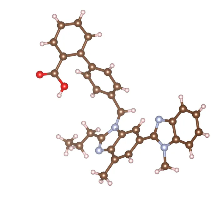
This shows the application example of XtaLAB Synergy-ED
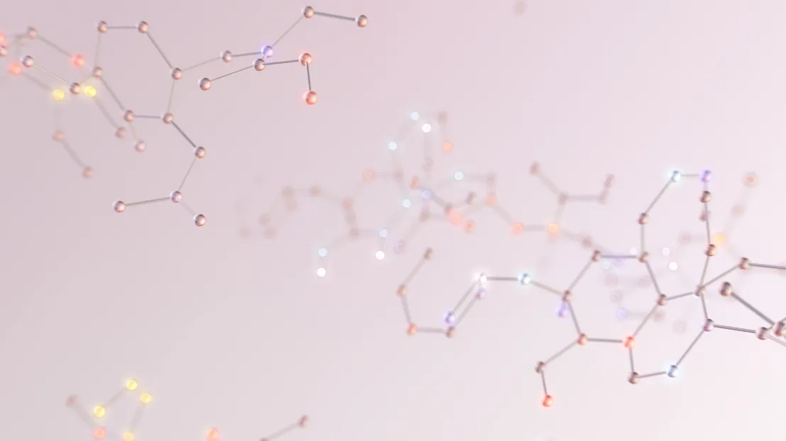
The molecule in the figure shows the 3D structure of Telmisartan (high blood pressure medicine). Just 16.7 micrograms of telmisartan was dissolved in 300μL ethanol and left at room temperature to allow the solution to gradually concentrate and form crystals. With XtaLAB Synergy-ED, a 3D structure was obtained with high accuracy.
From the above, XtaLAB Synergy-ED brings unprecedented value to a variety of drug discovery research scenes. Moreover, its structure analysis capability using a small sample volume is expected to be useful in the fields of drug discovery research as well as food, beverage, chemistry, forensic science, and omics.
Crystal Structure Analysis of New Functional Materials
New materials with various functions are being developed. These materials form the basis for the expected solutions to the issues the earth currently faces in many areas, including energy, resources issues, the development of science and technology, and medicine. It is no exaggeration to say that various functional materials are being utilized in a wide range of applications accompanied by the progress of nano technology, thus benefitting humans. It is very important to examine why these materials perform the functions that they do, and in particular, the crystal structures have a significant impact on their functions.
The techniques used to understand the crystal structure include X-ray diffraction system, NMR instrument, mass spectrometer, and electron microscopes. Among these, X-ray diffraction system, especially X-ray diffractometers for single crystal structure analysis are widely used as it allows for the precise determination of crystal structure. The sample size required for single crystal structure analysis using X-rays is considered to be 1 micron or more and measurement is difficult for functional materials that are smaller than this. Therefore, as a similar method for smaller single crystals, the single crystal structure analysis with the electron diffraction using electron beams instead of X-rays, has attracted attentions in recent years. This technique is called 3D ED/MicroED and is used to analyze structures of proteins and low-molecules, etc.
Different from X-rays, 3D ED/MicroED can acquire data that is possible to analyze from very small single crystals. In addition, the analysis technique used with X-rays can be applied as routine for analysis as is. Currently, many functional materials, in particular, catalysts, metal complexes, MOFs, COFs are often small crystals, and their structures used to be difficult to analyze. With the appearance of 3D ED/MicroED, there are high hopes for their structure analysis.
Synergy-ED is a dedicated 3D ED/MicroED system which can perform structure analysis of small single crystals. Data measurement as well as analysis routines are integrated, enabling seamless and automatic operations from measurement to data analysis. The number of papers and measurement examples are increasing, where the crystal structure from a small single crystal about several hundred nanomters is obtained, which is difficult to measure using X-ray diffraction. XtaLAB Synergy-ED is expected to be very promising and will be used for the analysis of functional materials of such minute size.